
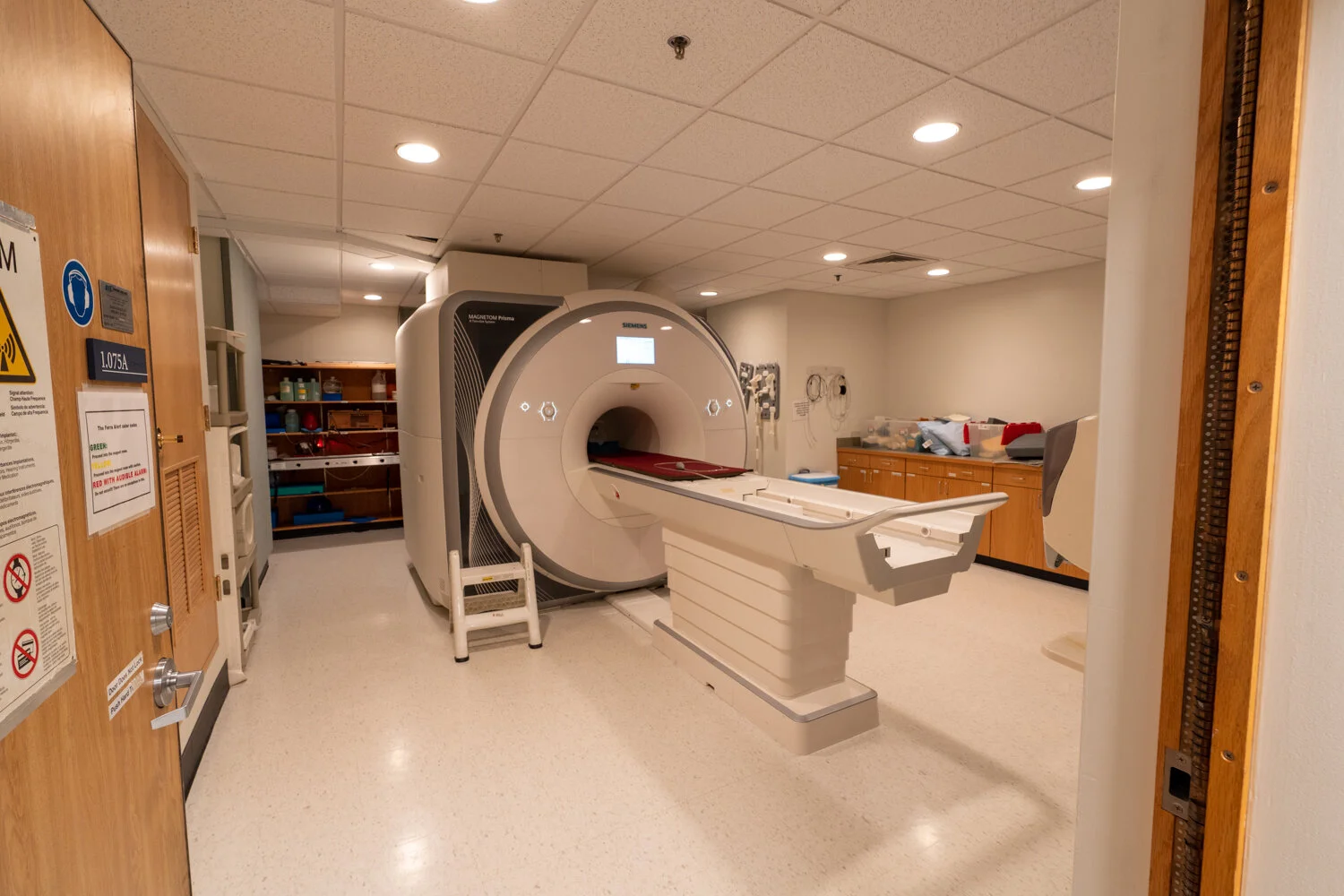
One of the more unique photos on my gallery page is a wide angle view of the bore (tube) inside of a new high-field (7-Tesla) MRI scanner at the research facility where I work (the MGH Martinos Center for Biomedical Imaging). I had a lot of fun making this photo happen and wanted to explain my process, as well as show some of the photos I took leading up to the final version.
The MGH Martinos Center is a research center in Charlestown, MA that is home to a variety of MRI machines used primarily for neuroscience and other biomedical research. It is part of Massachusetts General Hospital and is affiliated with Harvard Medical School.
Most pictures of MRI scanners are relatively boring, owing primarily to the fact that once they are active, the magnetic field is too strong to bring any kind of camera into the scanner room. If you search google images for “MRI scanner”, you get a wall of images similar to this one:
Picture I took of another scanner at the Martinos Center. This magnet is active so this is the closest anyone can bring a camera.
To take this picture, I first asked the Martinos Center safety personnel (Mary O’Hara) and the director of the center (Dr. Bruce Rosen) for permission to take my camera in to the magnet room before the magnetic field was at full strength. Thankfully I was given the all-clear to do this.
I went down to the scanner room after work one evening, and first took another instance of the typical, “boring” MRI photo. Note that since this was a brand new machine, there is still a lot of construction equipment around the room, the patient table was not complete, packing material is still present, etc. These photos were taken in August 2019, and the scanner room is now clinical-grade clean. We started to collect data on this in January 2020, shortly after it became operational.
Yawn…

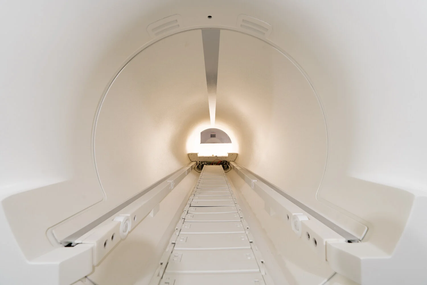
I started to take a few photos from different angles around the front of the scanner. I liked this center view but without a pro lighting setup it didn’t really pop.

Now I was starting to get some angles that would never have been possible under normal circumstances (with the magnet active).
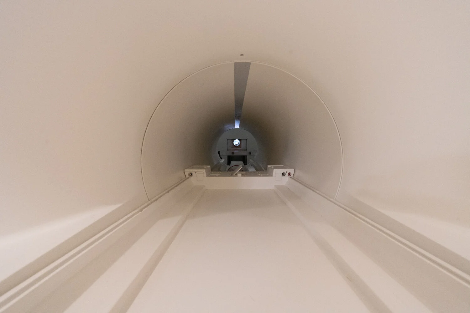
I wanted to capture some kind of “heading in” point of view, as if this is what you’d see if you were about to be scanned (not accurate because patients/volunteers would be looking straight up into a mirror…so I was taking some artistic license).

I made my way around the side of the scanner, and with the sliding compartment still open a lot of the electronic components were visible.
I went the rest of the way around, to the back of the scanner, where there was a little construction table at about the same height as the bore of the magnet. I did not have a tripod with me, so I placed the camera on this table and started framing this shot:
The same shot after some cropping and desaturation.
Here I used a longer exposure to get the light to start popping.

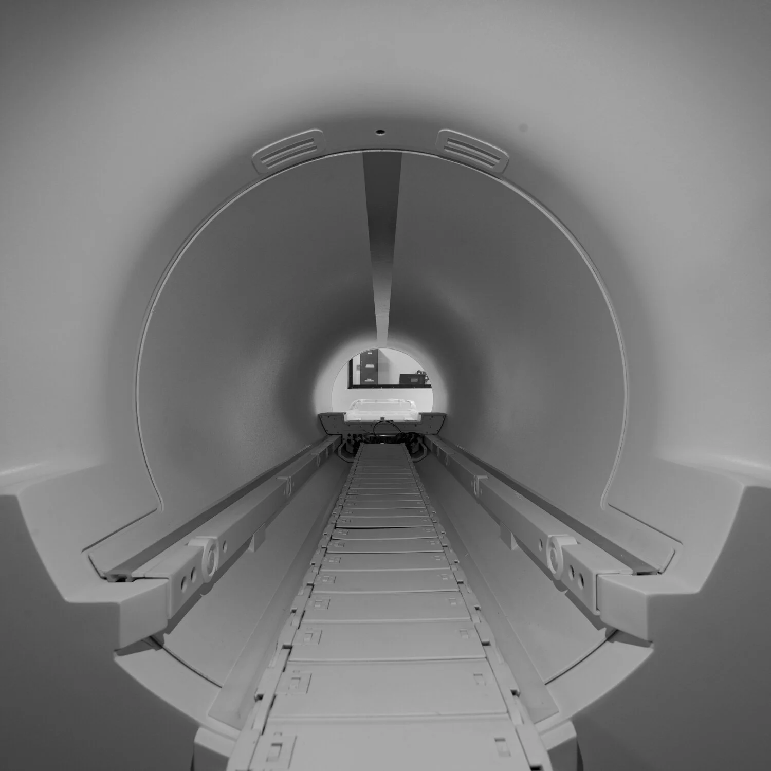
A grayscale version of the longer exposure shot.
I did like this perspective, but something about the bright lights in the room was still making me feel that this was just a plain image of an MRI tube - not something many people would be captivated by. I decided to turn all the lights off in the room and use only the light from my cell phone screen. I used a simple app (Screen Flashlight - for android) that could have the entire screen of my phone display a single color, which could be adjusted for color and luminance.

First image using my phone as a light source, with the screen glowing with a uniform green.

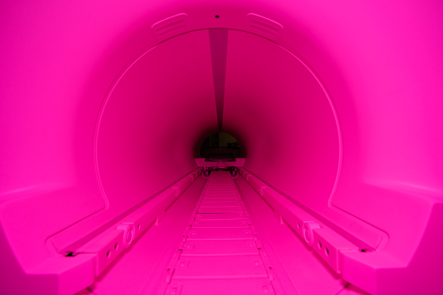
Magenta lighting a little intense…
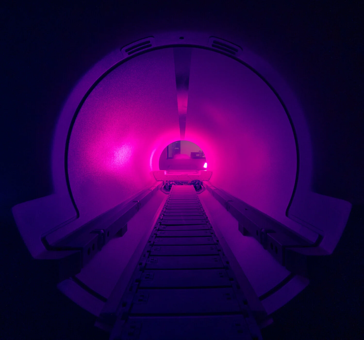
Whoa! Some colors were not working for this…
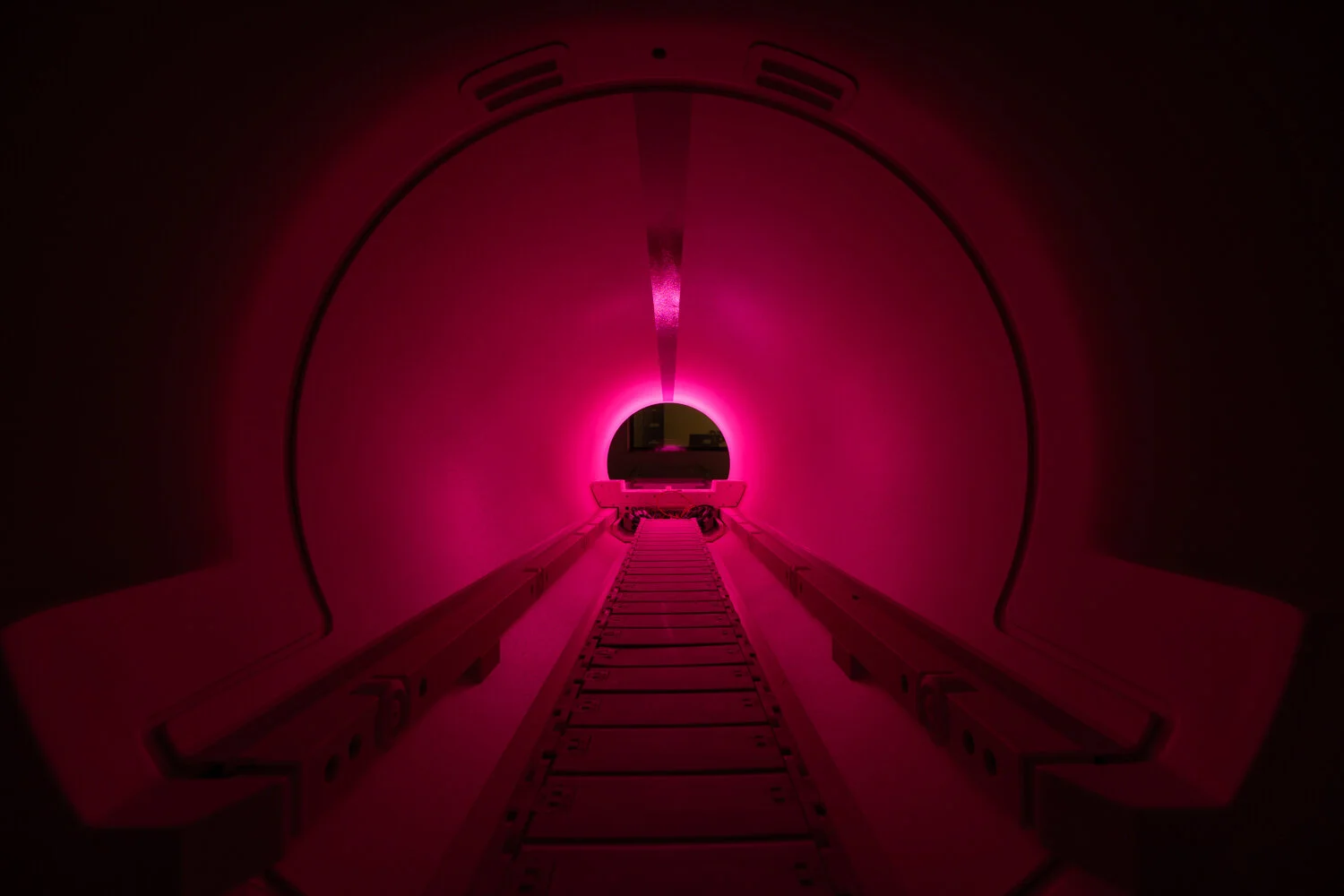
I then brought my phone around to the front of the scanner, and placed it to the side of the patient table at the threshold of the bore. I liked the effect this had, casting an eerie luminescence within the bore.
Cell phone screen is visible in this shot, just outside the bore on the right side.
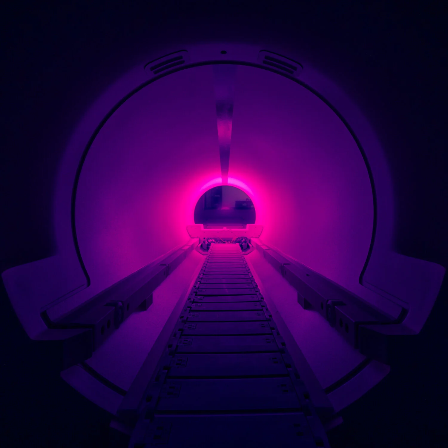
Here I adjusted the phone so it rested out of view while still casting its glow.
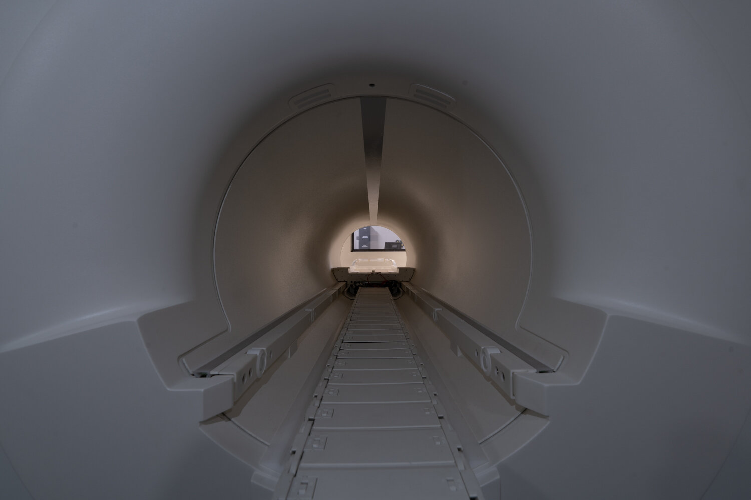
Although I really liked the perspective above, including the entire curvature on the bottom of the bore opening, I moved the camera a little closer and really like how this new perspective filled up the 3:2 dimensions of the entire camera sensor:
I took many photos using this same point of view, adjusting the colors on the phone app and playing with camera settings. With most exposures being 30s long, and the camera taking about 20s to process each of those, this part took something around 30 minutes before I felt I had a good enough variety of shots.
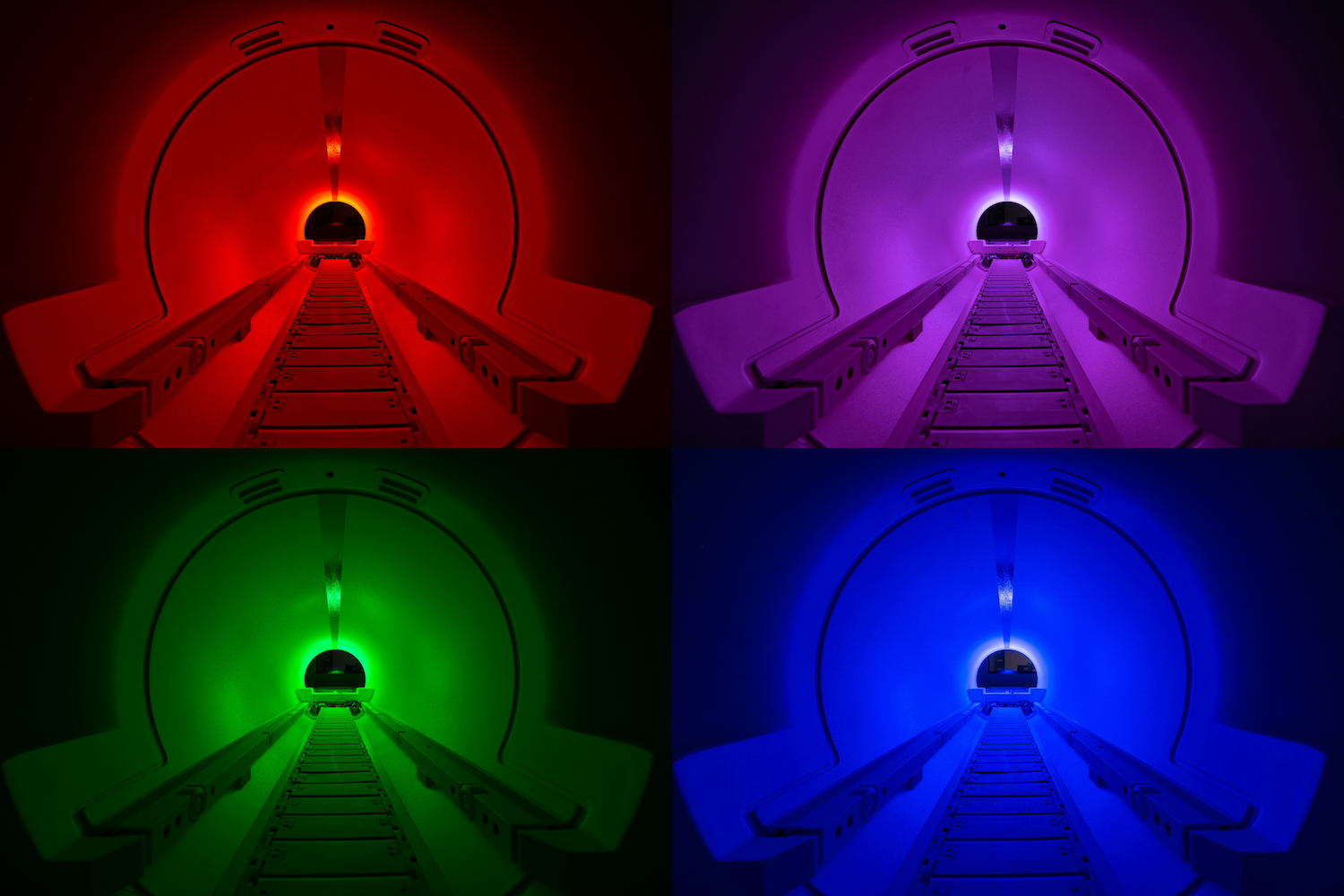
Various cell phone lighting colors I tried. No post-processing on these images - all colors were created in the room and these are the raw images.
It was hard for me to decide which of these color versions I liked more. I showed the images to a few coworkers at lunch and by consensus I was convinced the plain white light was the one that best demonstrated the features of the scanner, and made it the most apparent that this was in fact an MR scanner.
The camera settings for most of these final versions of the image were:
Sony Alpha 7 III camera body with Sony FE 16-35mm F2.8 GM lens.
30-second exposure
ISO 100
f/8 aperture
16mm focal length

The final image again.
My only intention with all of this was to get a unique photo that could only be obtained under these very temporary conditions (new scanner and magnetic field completely down). I thought I could share it with people at the Martinos Center and on Instagram (which I did). But then a couple months after taking this photo, MGH announced their annual image contest. Although many images submitted to this contest are taken with microscopes or other scientific imaging methods, it was open to any photography or images related to biomedical research at MGH. I submitted this photo (“Tunnel Vision”), along with another on my photography page (“One Four Nine”). It ended up being a finalist, and a printed version is now on display in the image contest exhibit in the atrium of the Martinos Center building.
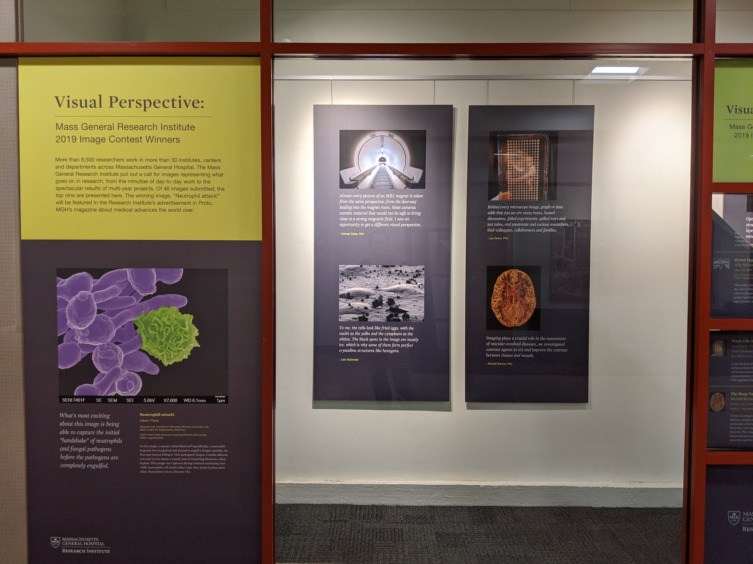
The MGH Image Contest finalist exhibit
This was a fun creative process for me so I was really glad it was well received.
As I mentioned earlier, one of the research studies I work on is using this magnet to collect data from patients who experience episodic migraines. The study is looking at changes in neurophysiology and autonomic nervous system functioning, and patients will participate in a multimodal treatment program that includes a combination of cognitive/behavioral stress-reduction training as well as vagus nerve stimulation. I hope to share some updates about that work in the future as well!